
Blog
Saving Limbs
Where NPWT Fits in the Diabetic Foot Ulcer Treatment Pathway
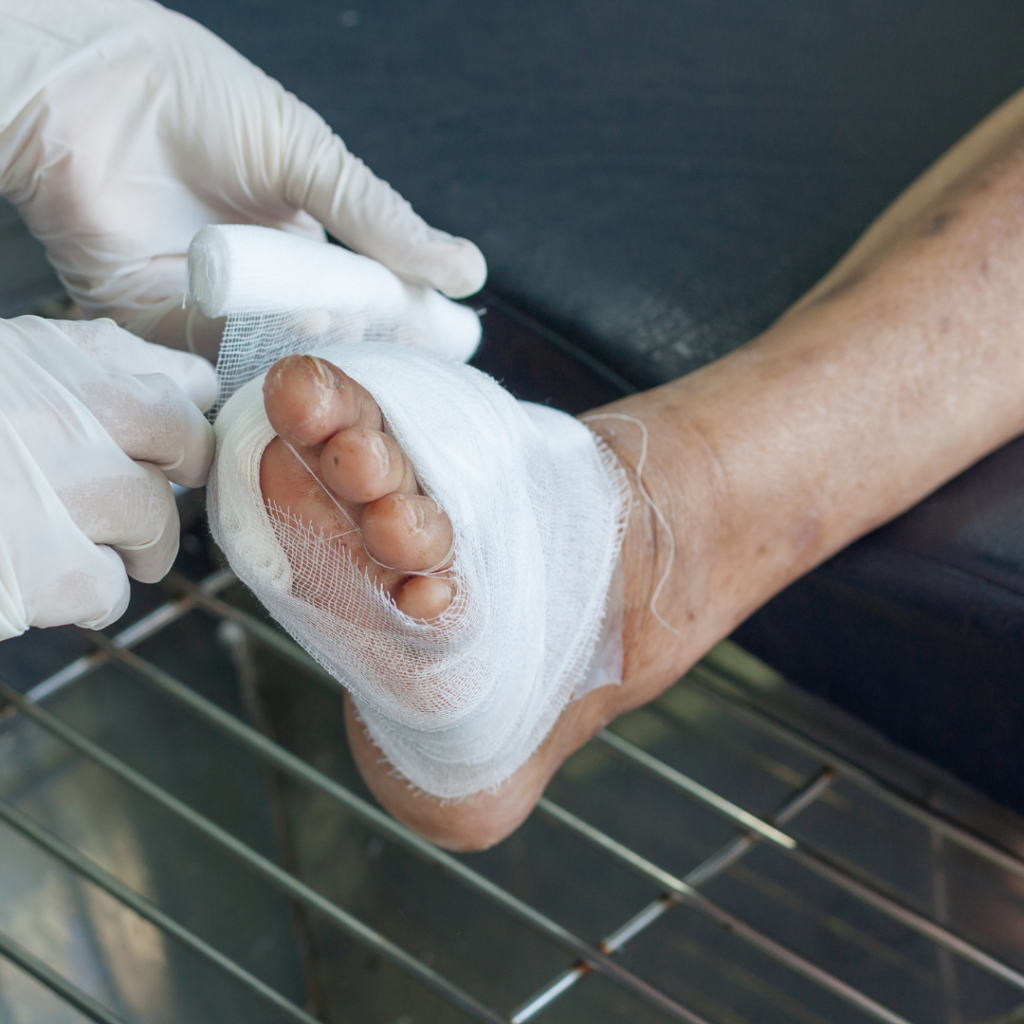
Every 20 seconds, someone somewhere loses a lower limb due to diabetes. Diabetic foot ulcers (DFUs) are among the most complex and serious wounds clinicians face. Without proper management, they are the leading cause of non-traumatic lower limb amputation.
This problem is widespread. The International Diabetes Federation reports that 589 million people had diabetes in 2024, and this number may reach 853 million by 2050.1 Each year, about 2–5% of patients develop DFUs, and up to one-third will have a foot ulcer at some point in their lives.2 The five-year mortality rate after a diabetes-related amputation is over 50%, which is similar to the survival rates of many cancers.2
The good news is that most cases can be managed to save the limb with a team approach. Negative pressure wound therapy (NPWT) has become an important tool in this process. While it is not a cure-all, NPWT helps address specific challenges in healing DFUs. This article explains where NPWT fits and why it matters.
Why DFUs Are Different From Other Wounds
Diabetic foot ulcers do not heal in a simple or predictable way. Three main problems often overlap and make them especially hard to treat:
- Peripheral neuropathy removes the pain that would normally alert patients to rest or seek help. Because of this, many DFUs go unnoticed until serious tissue damage has occurred. By the time the wound is found, it may be deeper, larger, or more infected than it appears.
- Peripheral arterial disease (PAD) reduces blood flow to the foot. Without enough blood, even well-managed wounds struggle to heal. Tissue lacking oxygen cannot form new tissue well, fights infection poorly, and may not respond to standard treatments. PAD affects nearly half of all DFU cases.2
- Infection is both common and serious in DFUs. Biofilm, deep tissue infection, and bone infection can all make treatment more difficult. Research in the Journal of Wound Care found that most chronic wounds contain biofilm, which keeps inflammation active, weakens the immune system, and increases the risk of amputation.3 In patients with poor blood flow and weak immunity, infections spread faster and are harder to control.
These three problems often occur together. For example, a patient with a foot ulcer on the sole may have all three at the same time, so treatment needs to address each issue.
What the Evidence Shows
The clinical evidence for NPWT in DFU management has grown substantially over the past two decades. A 2025 systematic review published in Diabetology analyzed 11 randomized controlled trials involving 1,699 patients and produced some of the most rigorous summary data to date.4
The key findings:
- Healing rates: In eight trials with 1,456 patients, NPWT made complete wound closure within 12 to 16 weeks about 46% more likely than standard moist wound care (risk ratio 1.46; 95% CI 1.21–1.76).4 The number needed to treat was about 8, which is significant given the seriousness of DFUs.
- Time to healing: Data from six trials showed that NPWT shortened the time to full healing by an average of 18 days compared to standard care (mean difference –18 days; 95% CI –28 to –8 days).4
- Granulation tissue formation: Five trials found that patients using NPWT were much more likely to reach 95% or more granulation tissue coverage, which is a key sign that the wound is ready for closure or grafting.4 There were also fewer minor amputations (risk ratio 0.71; 95% CI 0.50–1.00), though this finding is less certain and needs more research.4
- Hospital stay: Four trials reported that NPWT patients spent an average of 3.8 fewer days in the hospital, which can reduce both patient burden and care costs.4
- Adverse events: Even though NPWT provides more active wound management, it did not increase the rate of adverse events compared to standard care.4
A separate meta-analysis published in the World Journal of Diabetes (2025) covering studies from the United States, Chile, Pakistan, Italy, India, and Germany reached consistent conclusions: NPWT was associated with a significant improvement in wound healing rates (RR 1.46; 95% CI 1.22–1.76) and a significant reduction in amputation rates (RR 0.69; 95% CI 0.50–0.96).5 Granulation tissue formation time was reduced by an average of 19.54 days.5
Most RCTs in this area have required patients to have adequate blood flow, usually an ankle-brachial index of 0.7 or higher. So, these findings apply best to neuropathic or mixed-etiology DFUs with enough vascular supply. Clinicians should make sure revascularization is addressed before using NPWT in ulcers mainly caused by poor blood flow.4
The Multidisciplinary Framework: Where Does NPWT Enter?
Managing DFUs effectively is not the job of just one specialty. It usually takes coordination among wound care specialists, vascular surgeons, orthopedic or podiatric surgeons, infectious disease experts, and diabetes management teams. NPWT does not replace any of these roles, but it helps create conditions that make the rest of the treatment pathway more successful.
- After debridement, before closure: NPWT is usually started once the wound has been surgically cleaned and infection is controlled or being managed. At this point, the wound bed needs to be prepared for healing, and this is where NPWT is most helpful.
- During the granulation phase: NPWT helps form granulation tissue by keeping the wound moist, reducing swelling, and pulling the wound edges together. In DFUs, where healing is already difficult, this support can mean the difference between a wound that heals and one that does not.
- As a bridge to closure: For DFUs that may need skin grafting or surgical closure, NPWT prepares the wound bed by building strong granulation tissue. Without enough granulation, grafts are less likely to take, and closure is more likely to fail.
- Post-surgical stabilization. In some cases, especially after partial foot amputations or surgical removal of deeper tissue, NPWT is used to stabilize the remaining wound, manage exudate, and protect the surgical site during early healing.
The International Working Group on the Diabetic Foot (IWGDF) 2023 Wound-Healing Guideline gives a conditional recommendation for NPWT as an extra treatment for post-surgical diabetic foot wounds. It also stresses that NPWT should work alongside, not replace, best-practice measures like debridement, infection control, revascularization, and offloading.6
NPWT and Offloading: A Required Combination
No NPWT protocol for diabetic foot ulcers is complete without addressing offloading. Continued mechanical pressure on a plantar ulcer, even if it is well-dressed, will undermine healing no matter how effective the NPWT device is.
The current standard of care for neuropathic DFUs is total contact casting (TCC), which redistributes plantar pressure across the entire foot and eliminates focal loading at the wound site.6 Removable cast walkers (RCWs) and other offloading devices are also used, though adherence varies.
NPWT and offloading work together, not one after the other. While NPWT manages the wound environment, offloading addresses the mechanical cause of the wound. Both are needed. Clinicians using NPWT for DFUs should make sure there is a clear offloading plan and that the patient’s adherence is closely monitored.
For wounds on the bottom of the foot, using a wound vac device with an offloading system also means paying attention to how the dressing and tubing are set up to avoid pressure points or problems with the device.
Supporting Granulation Before Grafting
Skin grafting is one of the more common closure strategies for larger or deeper DFUs once the wound bed is adequately prepared. The quality of the granulation tissue bed is a primary determinant of graft success.
NPWT helps in this phase in two main ways. First, it speeds up granulation tissue formation by applying subatmospheric pressure that stimulates cell growth and new blood vessel formation.4 Second, it manages exudate, which in DFUs can be heavy and can damage the wound edges if not controlled.
A wound that reaches the grafting stage with a clean, well-vascularized, and exudate-controlled bed is much more likely to have a successful graft. On the other hand, wounds that go to grafting too soon, before enough granulation, often need repeat procedures, which increases treatment time and patient burden.
After grafting, NPWT is also used more often to keep the graft in place and reduce shear forces during the important early healing period. Clinical evidence supports this use, showing better graft take rates with negative pressure bolstering.7
Reducing Recurrence Risk
Amputation is not the only serious outcome in DFU care. Recurrence is also a major clinical problem. Research has found that three-year recurrence rates in patients with DFUs and chronic limb-threatening ischemia are similar to those seen in many cancers, showing the long-term risks of incomplete healing.2
NPWT helps reduce recurrence mainly by supporting strong wound closure. This means not just surface healing, but closure with enough granulation and protected tissue edges. Wounds that close with leftover sinus tracts, poor blood supply, or shallow healing are more likely to break down under the stress of walking.
Beyond the wound itself, reducing recurrence over the long term depends on patient education, changes in footwear, regular check-ups, and ongoing diabetes management. NPWT is just one part of a full limb preservation program, but lasting results need the whole system.
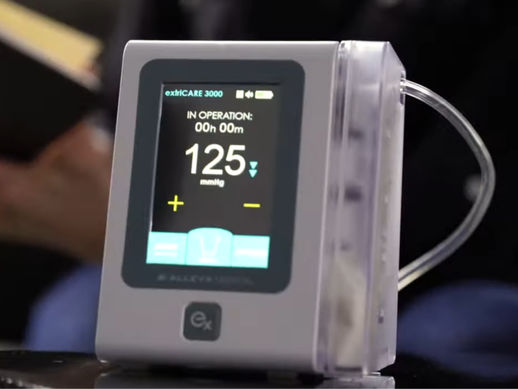
What to Look for in an NPWT device for DFU management
Not all NPWT platforms are equally suited to the clinical and logistical realities of DFU care. Several considerations are particularly relevant:
- Portability and wearability. DFU patients often manage wounds at home or in outpatient settings. A device that is small, quiet, and easy to use with daily activities helps patients stick to treatment and makes care transitions easier. The 2025 review in Diabetology found that single-use, ultraportable NPWT devices worked at least as well as traditional canister-based systems for complete healing (RR 1.57; 95% CI 1.22–2.03). This finding is important for home and community DFU care.4
- Dressing flexibility. DFUs vary in depth, shape, and location. NPWT systems that offer flexible dressing options, such as for tunneling wounds, undermined margins, or irregular wound beds, better meet the different needs clinicians see.
- Exudate management capacity. High-exudate DFUs require canister or dressing systems with sufficient capacity to manage output between dressing changes without leakage or device alarm fatigue.
- Integration with care transitions. Many DFU patients move between inpatient, outpatient, and home care. NPWT platforms that support continuity across these transitions, with consistent device interfaces, caregiver training, and clinical support, help reduce the risk of problems when care is handed off.
The Bottom Line for Clinicians
Diabetic foot ulcers are complex, high-risk wounds that need a coordinated, team-based approach. NPWT does not replace vascular assessment, infection management, or offloading, but within a structured treatment plan, it fills specific gaps that other treatments cannot address as well.
When used at the right stage, set up properly for the wound, and combined with offloading and diabetes management, NPWT supports granulation, wound bed preparation, and closure, making limb salvage possible. The evidence is clear: for well-perfused, neuropathic, and mixed-etiology DFUs, NPWT consistently does better than standard wound care in healing rate, time to closure, granulation quality, and lowering amputation risk.
For wound care teams managing DFU patients, the question is not whether to use NPWT, but how to use it most effectively within the overall treatment plan.
ExtriCARE provides NPWT solutions to help manage complex wounds in different care settings, including outpatient and home-based DFU care. Contact us to see how ExtriCARE can support your clinical team.
References
- Llanos S, Danilla S, Barajas-Nava L, et al. Negative pressure wound therapy for skin grafts and surgical wounds healing by primary intention. Cochrane Database of Systematic Reviews. 2018;(9):CD009261. https://doi.org/10.1002/14651858.CD009261.pub4
- International Diabetes Federation. IDF Diabetes Atlas, 11th ed. Brussels: IDF; 2024. https://diabetesatlas.org
- Armstrong DG, Boulton AJM, Bus SA. Diabetic foot ulcers and their recurrence. New England Journal of Medicine. 2017;376(24):2367–2375. https://doi.org/10.1056/NEJMra1615439
- Malone M, Bjarnsholt T, McBain AJ, et al. The prevalence of biofilms in chronic wounds: a systematic review and meta-analysis of published data. Journal of Wound Care. 2017;26(1):20–25. https://doi.org/10.12968/jowc.2017.26.1.20
- Theodorakopoulos G, Armstrong DG. Negative-pressure wound therapy in diabetic foot management: synthesis of international randomized evidence over two decades. Diabetology. 2025;6(11):126. https://doi.org/10.3390/diabetology6110126
- Efficacy and safety of negative pressure wound therapy for the treatment of diabetic foot ulcers: a meta-analysis. World Journal of Diabetes. 2025;16(6):103520. https://www.wjgnet.com/1948-9358/full/v16/i6/103520.htm
- Chen P, Vilorio NC, Dhatariya K, et al. Guidelines on interventions to enhance healing of foot ulcers in people with diabetes (IWGDF 2023 update). Diabetes/Metabolism Research and Reviews. 2024;40(3):e3730. https://doi.org/10.1002/dmrr.3730
